Model systems and specialised techniques
Main content
Xenograft models
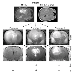
In our laboratory, patient GBM biopsies that are minimally propagated in vitro as spheroids are serially implanted intracranially into athymic rats to develop xenograft tumours that closely resemble the original patient biopsy. As opposed to the well circumscribed, non-invasive tumours established from GBM cell-lines, xenografts established from tumour biopsy-spheroids show striking histological and genetic features of the patient GBMs in situ. By successive passaging in vivo, invasive non-angiogenic tumours may be established (Phenotype I), invasive and angiogenic (phenotype II) and non-invasive angiogenic, (phenotype III), Fig 1. These models are important for assessing therapeutic efficacy of novel compounds in a heterogeneous tumour and investigating the relevance of bio-markers in tumour progression. The University of Bergen has state-of-the-art molecular imaging platforms including a high resolution 7 Tesla animal MRI machine, in vivo optical bio-imagers for drug delivery and bio-distribution. We have numerous animal breeding and husbandry facilities for preclinical studies.
Patient Biobanks
Our studies also utilise a large, clinically annotated patient GBM tissue, and blood biobank with continual inclusion. Biobanks are useful research tools for directly identifying biomarkers involved in malignant progression that may aid diagnosis, development of novel therapies and extend our understanding of basic mechanisms of disease processes.