Åpningssymposium for det nye Hyperion Imaging System
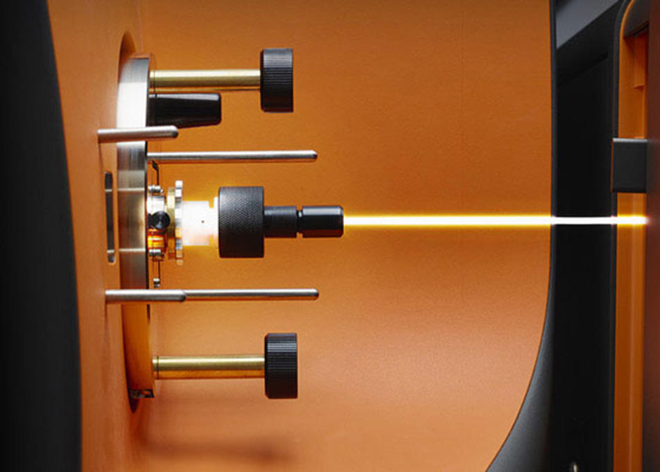
Centre for Cancer Biomarkers CCBIO og kjernefasiliteten Bergen Flow Cytometry har gleden av å presentere det nyerhvervede Hyperion Imaging System, som nå er tilgjengelig for forskergrupper ved det Medisinske fakultet. Åpningssymposiet vil presentere nye forskningsmuligheter som Hyperion Imaging System åpner opp for, gi eksempler på banebrytende forskning som Hyperion har lagt til rette for, og informere om de praktiske prosedyrene angående tilgang til maskinen og bruken av den.

Hovedinnhold
The Centre for Cancer Biomarkers CCBIO and the Bergen Flow Cytometry Core Facility are happy to introduce the Hyperion Imaging System to our research community – surpassing the inherent limitations of fluorescence detection by using pure metal tags that are separated by mass instead of by wavelength.
This is next generation immunohistochemistry: explore tissue biology with 30 antibodies simultaneously!
Setting a new standard in highly multiplexed protein detection, the system will enable our researchers to simultaneously detect up to 37 markers from a single tissue section by IMC. This allows for a comprehensive analysis of cellular phenotypes and their interrelationships within the spatial context of the tissue microenvironment.
The opening symposium will introduce you to the research possibilities offered by the Hyperion Imaging System and give examples of groundbreaking research in a high level keynote lecture.
Leigh-Anne McDuffus from the Fluidigm Corporation will explain the features and possibilities of the Hyperion Imaging System. Keynote speaker will be Dr. Dario Bressan, Head of the IMAXT Laboratory, CRUK Cambridge Institute, University of Cambridge, UK.
All participants are also invited to an informal interaction following the symposium outside of the auditorium, with delicious tapas.
Who: Everybody; researchers and technicians, PhD fellows and postdocs, students, guests and administrative staff.
Place: Auditorium 1, BBB
Registration: please use this link: https://skjemaker.app.uib.no/view.php?id=6031041
To make sure we order enough tapas, we prefer if you register as soon as possible and within January 31st.
Tentative program
13.00 Introduction
13.15 The Core Facility Concept & Procedures for Access
13.30 The Hyperion Imaging System: Leigh-Anne McDuffus, Fluidigm Corporation
14.15 Keynote lecture: Dr. Dario Bressan, University of Cambridge
15.15 Mingling session with tapas
Chairperson: Sonia Gavasso
Abstract
Abstract for Dr. Dario Bressan's lecture:
Building and exploring molecularly annotated 3D-tissue atlases to understand tumour heterogeneity and micro-environment
It is becoming increasingly obvious that tumours cannot be properly studied in depth without considering the tightly woven network of relationships between them and their surrounding tissues, the so-called tumour micro-environment. In a naturally occurring disease, different clones of cancer cells are constantly mutating and interacting with each other and with cells forming stromal components like fibroblasts, immune cells, vessel endothelium, and others. These interactions have a huge role in the progression of each tumour and its ability to recur and resist therapy.
While the advent of single cell “omics” technologies is allowing us to look at the micro-environment with an unprecedented amount of detail, these are still operating mostly on dissociated tissues, and are therefore lacking spatial information. This is a huge limitation, since the tumour micro-environment is clearly spatially organized, and many of its properties are likely resulting of its local structure.
Our laboratory, together with many others forming the IMAXT project consortium, is optimizing and integrating a series of in-situ methods to produce tri-dimensional, molecularly annotated atlases of tumours combining single cell genomic, transcriptomic and proteomic information; these include high-throughput imaging and sectioning, imaging mass cytometry, in situ sequencing and multiplexed in-situ hybridization. Specifically, we are applying these methods to breast cancer using a combination of animal models, patient-derived xenografts and human samples. This is providing insights on phenomena like immune re-modeling, vascular mimicry and the acquisition of intra-tumour heterogeneity, which are of high relevance in shaping cancer progression.
Biography – Dr. Dario Bressan
Dario Bressan graduated in Molecular Biology and Neurobiology from the Scuola Normale Superiore of Pisa, Italy, in 2008. He then moved to the USA, where he obtained a PhD from Cold Spring Harbor Laboratory, NY, working with Dr Greg Hannon.
During his Ph.D he worked on brain connectivity mapping, and developed tools to use light as a “tag” to selectively purify and analyse specific parts of a tissue, allowing the measurement of the gene expression of these components with high resolution without the need for microdissection.
Since 2014, Dario has been a research associate at the CRUK Cambridge Institute developing tools that eventually became part of the IMAXT project, a large endeavour aimed at producing tridimensional, multi-parametric maps of tumours to understand tumour clonality and micro-environment which received one of the first 20 million “Grand Challenge” grants from Cancer Research UK.
He is now heading the IMAXT laboratory, an interdisciplinary group of researchers at the CRUK Cambridge Institute focused on implementing and applying at scale a series of technologies aimed at the production of tri-dimensional, data-rich maps of breast tumours, which are going to be used to improve our knowledge of how cancer evolves and interacts with its host.
