Leica Stellaris
Hovedinnhold
Room 6C120dA
Telephone 555 86456
Responsible contact persons endy.spriet@uib.no and hege.dale@uib.no.
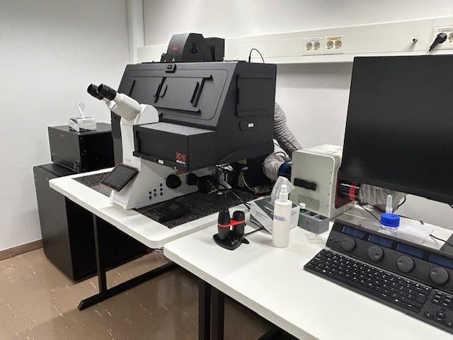
Leica's next-generation Stellaris was launched in November 2024 and installed at MIC in March 2025. Compared to our existing Leica confocal microscope, it features several exciting new advancements, highlighted in the first four points below. The microscope is equipped with five Power HyD detectors (the maximum number) and a white light laser (WLL) with a flexible excitation range from 440 to 790 nm. Additionally, it includes a camera and 10x and 20x air objectives for acquiring fast overview images.
- WLL (440-790 nm): The range of fluorescent dyes can now be extended into the near-infrared (NIR) range, which expands the panel of possible targets while maintaining good spectral separation of the fluorophores.
- Spectraplex software package: Facilitates the setup, acquisition, and analysis of up to 15 fluorophores simultaneously within the same sample.
- Fluorescent camera: Enabeling fast overviews using the Navigator software package which is particularly useful for examining tissue sections and identifying relevant areas without the need to search through the ocular. It is also valuable for detecting rare events in cell cultures. The Navigator also facilitates efficient sampling by allowing the setup of multiple z-scans in different positions, which can be acquired sequentially without requiring anyone to be present.
- TauScence: Fluorescence lifetime-based information, which can be used to minimize unwanted autofluorescent background in tissue samples, separate fluorescent dyes with overlapping spectra, and perform biofunctional analysis.
- Lightning software package: For high resolution imaging and image enhancement by adaptive deconvolution.
- Resonant scanner: For fast live cell imgaing (low light exposure) with the possibility for dynamic signal enhancement (DSE) to enhance the temporal dynamics and overall image quality.
The system is also equipped with:
- Tokai Hit Cage Inkubatorsystem for temperature (370C) and CO2-controll for live cell imaging.
- Auto Focus Control.
- Motorized (Super Z Galvo Stage Type H) which allows tile scan and multipoint acquisition.
Lasers / Laser lines:
- Blue diode (BD) - 50 mW 405 nm
- White Laser (WL) - lambda lange 440-780 nm, freely tunable in 1 nm steps, power approx. 1.5 mW per line, pulsed
Objectives:
M | NA | Type | IM | WD (mm) | Max FOV (um2) |
5x | 0.15 | HC PL Fluotar | Air | 13.7 |
|
10x | 0.40 | HC PL APO CS2 | Air | 2.56 |
|
20x | 0.75 | HC PL APO CS2 | Air | 0.62 |
|
20x | 0.75 | HC PL APO CS2 IMM | Water/Glyc/Oil | 0.680 | 775x775 |
40x | 1.25 | HC PL Apo CS2 | Glyc Corr | 0.35 |
|
40x | 1.10 | HC PL APO CS2 | Water MotCorr | 0.62 |
|
63x | 1.4 | HC PL APO CS2 | Oil | 0.14 |
|
M: Magnification / NA: Numerical aperture / IM: Immersion medium / WD: Working distance / FOV: Field of view