Mediso nanoScanPC PET/CT
Hovedinnhold
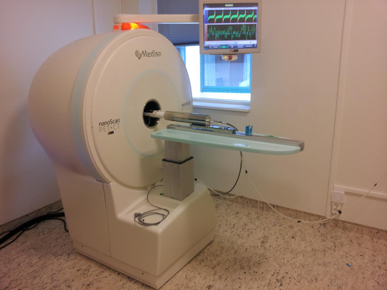
The nanoScan PET/CT from Mediso (Mediso Ltd) is mainly for mice and rats (or animals with similar size) offering high resolution (0.7 mm PET and 0.02 mm CT) and high sensitivity imaging. We have, in close collaboration with the PET center at Haukeland University Hospital, an on-site cyclotron and a fully equipped radiochemistry lab for production of a wide range of PET tracers/radionuclides (see table below). We also have a gamma counter (Wizard, 2480) available for use.
Adjacent to the scanner, we have a dedicated animal room for keeping animals during short- or long-term projects. Personnel from the animal facility organize the use. This also includes animal transport between the different departments if needed.
MIC can support you in all parts of the project; including planning, scanning, image post-processing and analysis.
Room: 7921, floor 0 at Center for nuclear medicine/PET, Parkbygget, Haukeland University Hospital.
Responsible MIC personnel: Hans Olav Rolfsnes (Hans.Rolfsnes@uib.no, phone 55 58 65 81)
Responsible animal technician: Helen Otterå (helen.otteraa@uib.no)
Click here for ANIMAL HOUSING FACILITY and ANIMAL TRANSPORT FORM
PET (positron emission tomography) an invaluable non-invasive diagnostic and research tool that provides 3D images and quantitative parameters of perfusion, proliferation and/or metabolic activity of tissue. These images result from the use of different substances of biological interest (sugars, amino acids, metabolic precursors, antibodies etc.) labeled with positron emitting radioisotopes (radionuclides). The technology is developing rapidly, resulting in a number of new tracers being developed and introduced regularly.
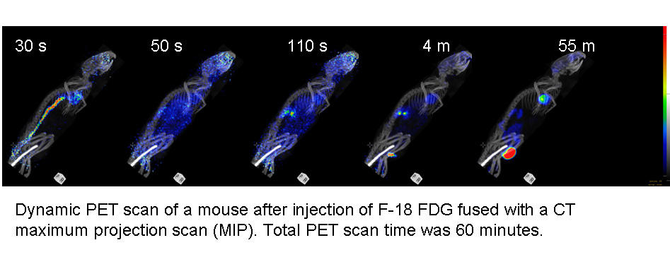
The most commonly used PET tracer is 18F-FDG (fluorodeoxyglucose), also called radiolabelled glucose. This tracer is used in most PET centers worldwide, and is an imperative tool in clinical cancer diagnosis, staging and management. FDG is an analogue of glucose and is taken up by living cells via the first stages of the glycolytic pathway. The rationale behind its use as a tracer is based on the increased glucose uptake by tumor cells. FDG uptake in tumors may correlate with tumor growth and viability, thus metabolic quantification from PET images may provide useful information regarding tumor characterization and treatment response. Other tracers MIC use at a regular basis include FMISO, which is a marker for hypoxia and FLT, which is taken up by proliferating cells. Below lists the tracers currently available and previously used, however, additional tracers may be produced by appointment.
Nuclide | Tracer | Application |
18F | FDG (fluorodeoxyglucose) | Glucose uptake eg. distant metastasis detection |
18F | FLT (fluorothymidine) | Cell proliferation eg. tumor cells |
18F | FMISO (fluoromisonidazole) | Hypoxia eg. tumor hypoxia |
18F | FET (fluoro-ethyl-tyrosine) | Amino acid metabolism eg. glioma/brain tumors |
18F | PSMA (prostate-specific membrane antigen) | Prostate cancer detection (coming 2018) |
18F | Choline (fluoromethylcholine) | Phospholipids metabolism eg. prostate cancer |
18F | NaF (sodium fluoride) | Bone scanning eg. bone regeneration and bone metastasis detection |
18F | DOPA (hydroxyphenylalanine) | Pancreatic hyperinsulinism |
11C | Methionine (methylmethionine) | Amino acid metabolism eg. glioma/brain tumors |
68Ga | DOTATOC | Somatostatin receptor expression, detection of neuroendocrine carcinomas (coming 2018) |
We have equipment and personnel to label your compound of interest: Please inquire | ||
I-124 | (your taget) | Eg. I-124-peptide/nanoparticle/protein |
Zr-89 | (your target) | Eg. Zr-89-antibody (bevacizumab/avastin has been carried out previously) |
CT (computer tomography): Provides anatomical information about the tissue of interest and by combining PET and CT, both functional and anatomical information can be provided. CT can be used in combination with PET, or as a standalone tool. We can also produce the sodium fluoride tracer NaF, for bone imaging. Combining CT with NaF-PET can be a useful for bone regeneration research.
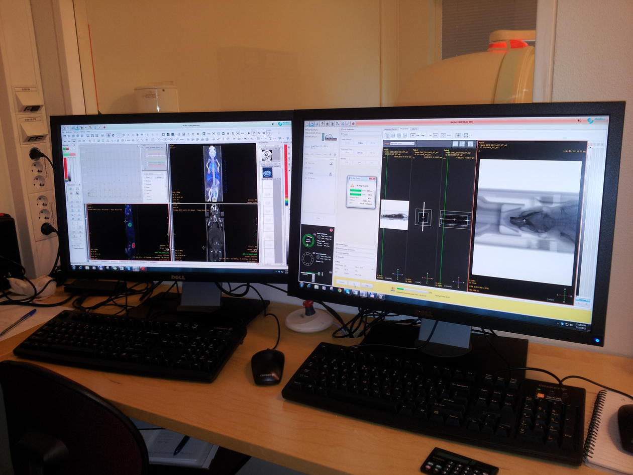
Image analysis: we have two dedicated computers available for users, where one is located in the scanner room and the other is located in the data analysis room at the 6th floor at BBB. We have the integrated software from Mediso “InterViewFusion” for analyses, in addition to PMOD (Version 3.8) for more advanced (kinetic) analyses.
Please contact Heidi or Helen for further information or a tour of the facilities.