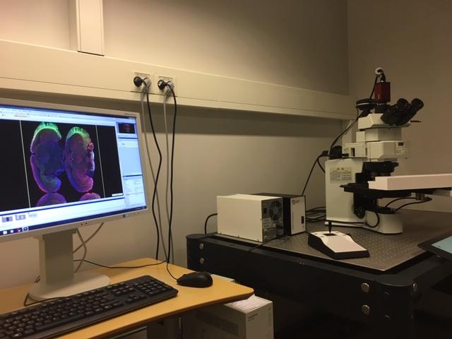
Olympus VS120 S6 Slide scanner
Main content
Olympus VS120 S6 Slide scanner is able to scan slides with tissue or cells either with brightfield or widefield fluorescence, in an automated or manual mode. The images are created as seamless tile scans of many small images over the field of interest. You can for example combine a large overview at low magnification with layers of chosen regions at higher magnification, and all images belonging to one slide is saved in the same raw data file. For viewing and analysing we recommend the freeware QuPath (see below).
Key features:
- Own software module facilitating scanning of slides with tissue microarray (TMA) in brightfield.
- Upright microscope that can fit up to 6 slides simmultanously.
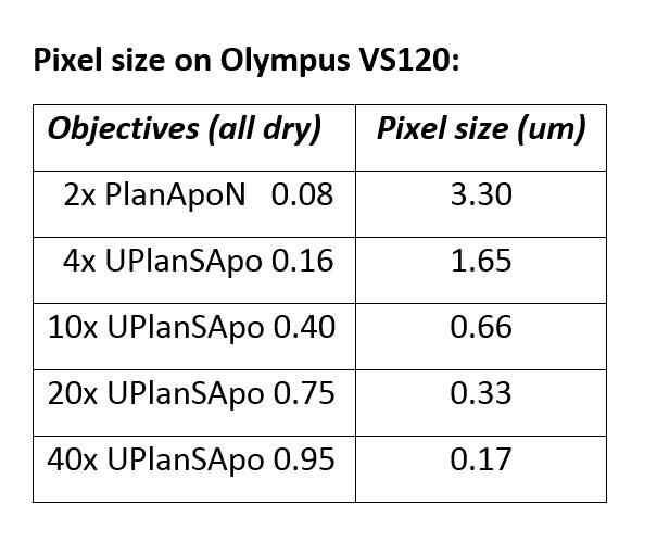
- 5 air objectives, maginifcation 2x, 4x, 10x, 20x, and 40x (see details and pixel size in table to the right).
- Fluorescence filters: blue, green, red, and far red (see details in table to the right).
- B/W camera for fluorescence: Hamamatsu ORCA-Flash 4.0 V3 (2048x2048 px, 6.5 um2)
- Color camera for brightfield: Allied Vision Pike F-505 (2452v2054 px, 3.45 um2)
Viewing and analysing data:
QuPath: A powerful freeware that opens VSI-files (raw format from the slide scanner). It is compatible with Windows, Mac OS X and Linux. This is a software under continous development, so make sure that you occasionally check for updates.
- Installation and information Welcome to QuPath!
- System requirements.
- "Speaks" with Fiji (ImageJ) for easy transfer of images both ways.
- Tutorials on YouTube recommended for first time users.
- Webinar "Quantitative Pathology & BioImage Analysis: QuPath" 29 April, 2020.
- Important: Remember to cite the software in your work!
Fiji (ImageJ): VSI-files can be opened via the OlympusViewer plugin, which can be downloaded here. Follow the description for installation and opening of files (restart required for functionallity).
OlyVia: Olympus own free program for viewing. It can be downloaded under Virtual Slide Microscopes, or you can get a copy from MIC (not MAC compatible).


