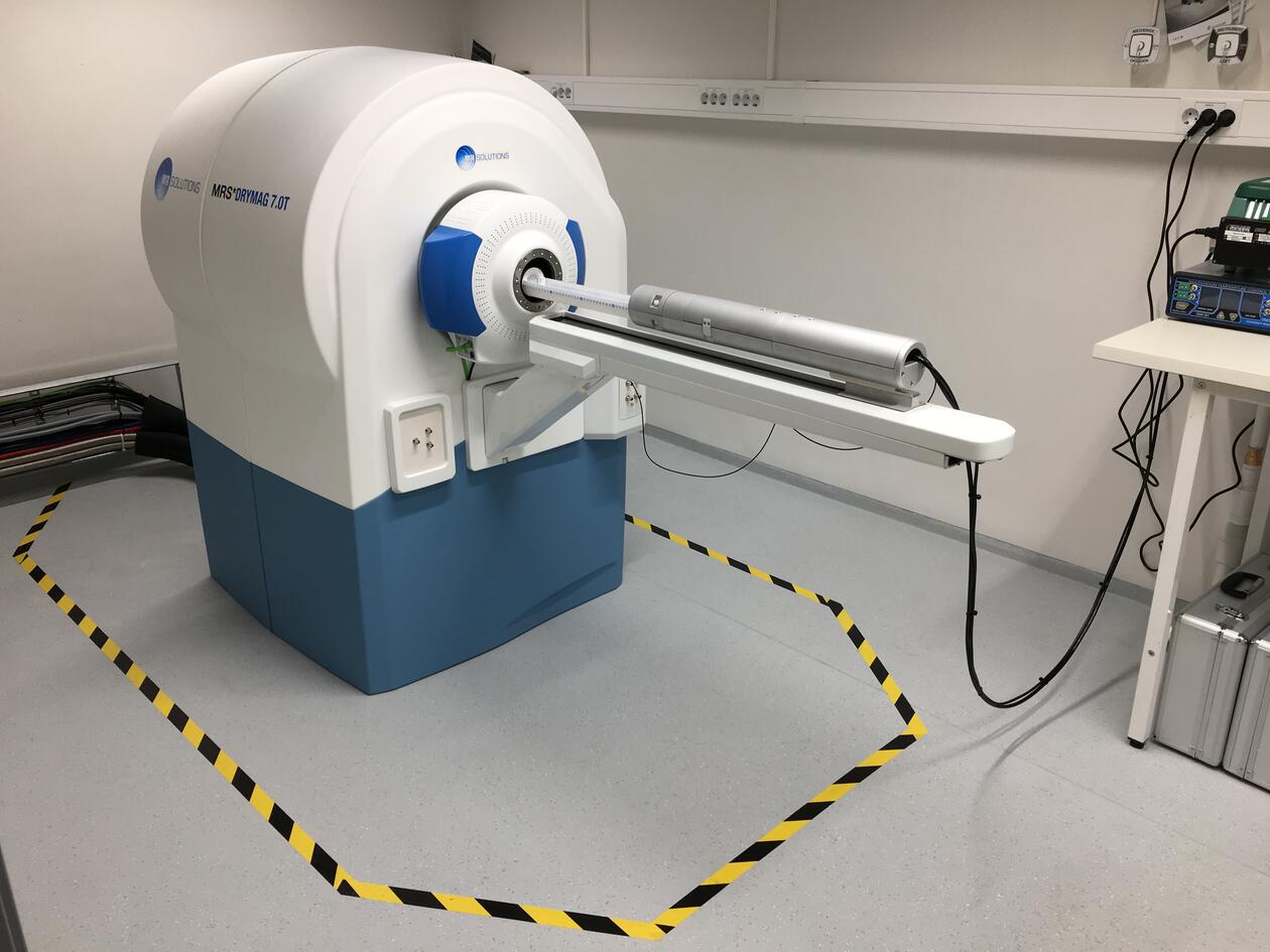
MR Solutions Powerscan 7T PET/MR
Main content
Room 3033/3034, Vivarium (3rd floor).
The PET/MR system is located adjacent to the optical imaging equipment, and ultrasound equipment is available next door, which makes it easy to combine different imaging modalities.
Responsible contact person: Hans Olav Rolfsnes
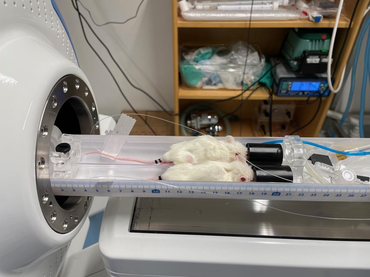
The system (installed April 2021) can perform in-vivo MR and PET imaging and MR spectroscopy of small animals, typically mice and rats but we can also image e.g. euthanised zebrafish. It consists of a 7T MR scanner and a 3 ring (15 cm axial field of view) PET scanner, which can perform MR imaging alone or sequential MR and PET imaging where the MR images serve as anatomical reference for the PET images.
For more details about PET imaging read here.
Examples of available imaging and spectroscopy techniques:
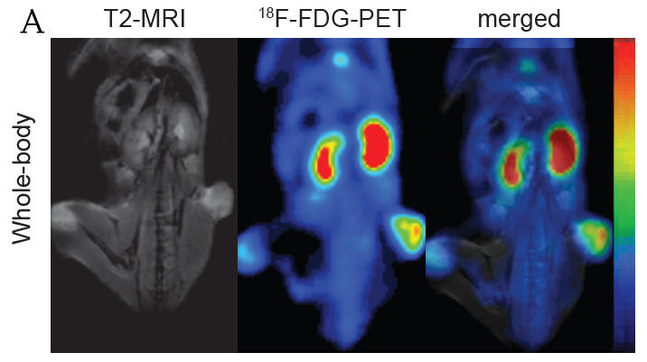
- PET/MR imaging (ex. picture A).
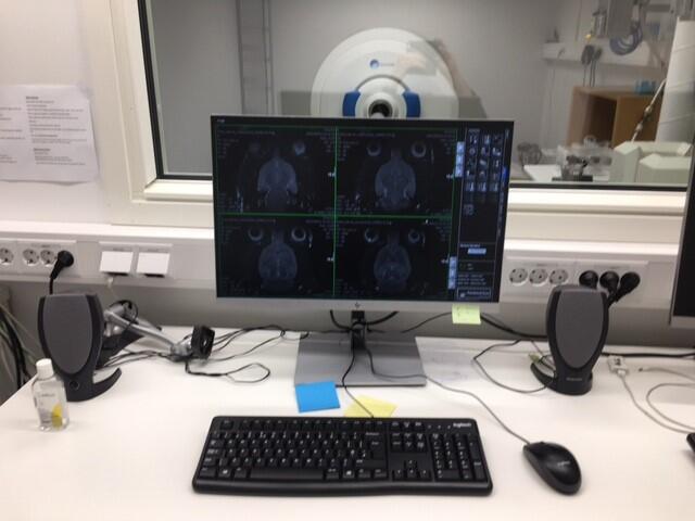
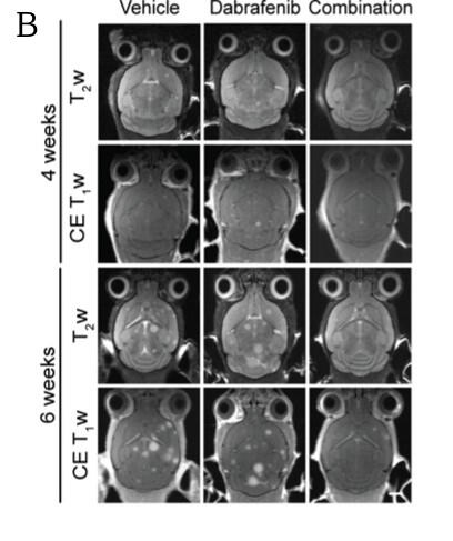
- Anatomical imaging such as standard T1 and T2 weighted images (ex. picture B).
- Diffusion weighted imaging (DWI): used to investigate changes in diffusion or apparent diffusion coefficient (ADC), e.g. to provide information about cell density in a tumour.
- Diffusion tensor imaging (DTI): typically used to investigate fibre pathways in the brain (or spinal cord) where diffusion is highly directional. May for example be used to study deformation and integrity of white matter fibre tracts.
- Perfusion imaging (DCE): used to study the rate of blood flow through tissue or organs to provide information about pathology, metabolism etc. An example is to investigate the integrity of the blood-brain barrier.
- Relaxometry: monitor/map T1, T2 and T2* relaxation times to characterise tissue properties, e.g. to differentiate between normal and abnormal (cancerous) tissue.
- Angiography: time-of-flight measurements to visualise the vascular structure and to quantify blood flow in an area.
- Cardiac imaging.
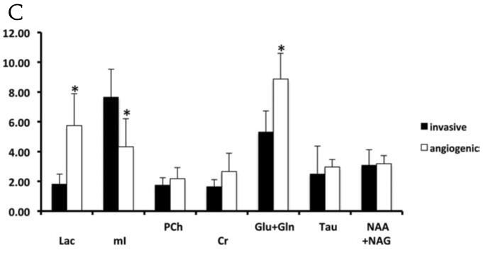
- MR spectroscopy (PRESS, MEGA-PRESS etc): used for looking at the presence of various metabolites and whether they have been up- or downregulated (ex. picture C).
Technical details of the system:
- System: Powerscan 7T PET/MR PET CLIP-ON 803.
- PET system: 15 cm axial field-of-view, up to 0.7 mm resolution.
- MR system: Magnetic field strength: 7T (297 MHZ for 1H).
- RF volume coils (inner diameter):
- mouse head (20 mm)
- mouse body (38 mm)
- mouse hotel for up to 3 mice (65 mm)
- rat head: (42 mm)
- rat body: (65 mm)
- RF surface coils for mouse head and rat head imaging.
- Gradient strength: 600 mT/m.
- Dedicated animal beds with built-in air heating.
- Life monitoring system for respiratory and temperature monitoring.