Optical Imaging.
Main content
Room 3030, 3rd floor Vivarium, HUS
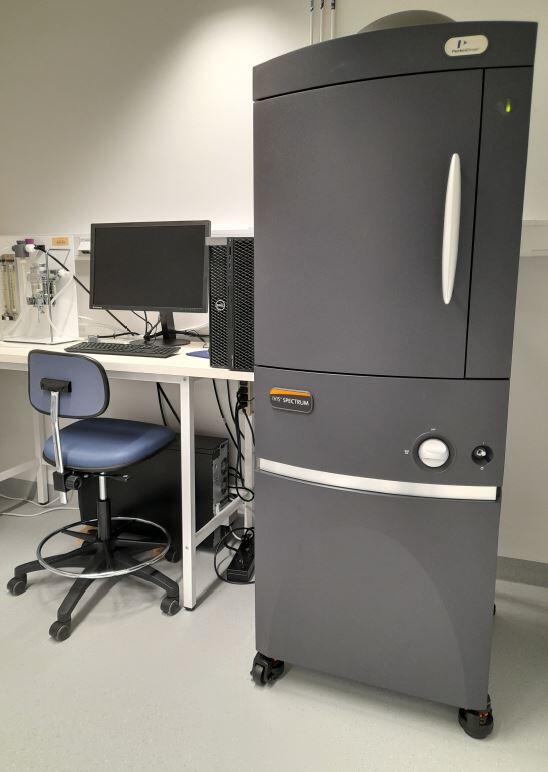
IVIS Spectrum is an optical imaging system for small animals. The system detects photons in the visible range and is highly sensitive. The imager has space for 5 mice simultanously and each scan can take up to a minute, maximizing the amount of specimens in pre-clinical studies.
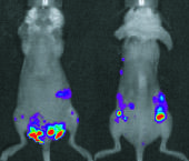
Optical imaging is based on detection of fluorescence and/or bioluminescence signal. It is a minimal- to non-invasive technique that enables the visualization of cancer cells in vivo, and can be used to follow disease progression in the animals over time to quantify the cancer burden. Bioluminescence imaging (BLI) is based on the expression of luciferase reporter gene in engineered cells.
Responsible contact person is Emmet Mc Cormack.
• Specifications
Model/Supplier: IVIS Spectrum In Vivo Imaging System (PerkinElmer)
Application: In vivo detection of near IR and GFP/Luminescent probes
| System | Excitation | Emission | Application |
| IVIS | 400-760 | 500-850 | Bioluminescence & Fluorescence |
• Links