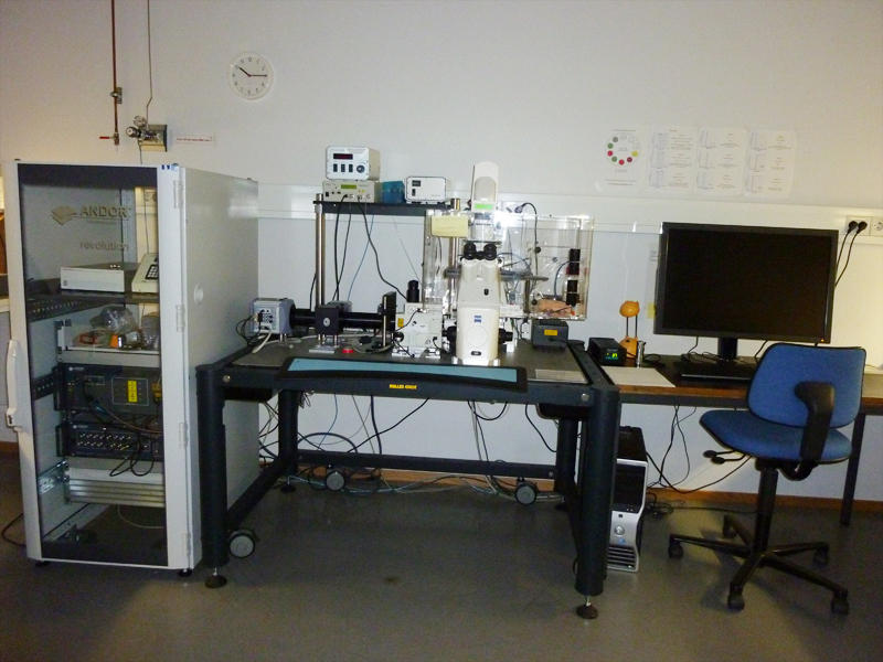
Andor Revolution Imaging System (Spinning disk)
Main content
Room 6C129bA
Telephone 555 86879
This instrument is ideal for rapid live cell imaging (movie example 1) and allows you to observe the entire process of interest from start to finish with less phototoxicity and bleaching. It's also possible to look at larger structures, see movie example 2 where a calcium flux is observed after stimulation of a dissected live blood vessel.
The excitation light passes through a spinning disk containing an array of pinholes and is focused onto the specimen through the objective of the microscope. Emitted light passes back through the same pinholes which results in exclusion of the emmission outside the focal plane. The light from the focal plane is detected by a very sensitive camera and a confocal image is therby generated in real time, which means approx. 30 frames per second. The system is sensitive and fast but the acquisition speed is still dependent on how strong the fluorescent signal is.
Responsable contact person is Hege Avsnes Dale
• Specifications
The system comes with a Zeiss Axiovert (model 200) microscope and is equipped with a heating chamber and a CO2 controller.
The equipment has a Yokogawa CSU-21 confocal scanning unit and a iXonEM+ DU-887 EMCCD camera (16 bit / 512x512). If needed, it is also possible to use a Hamamatsu Ocra ER Firewire CCD camera (12 bit digital / 1344x1024 pixels).
Laser lines (nm):
488, 561, 640
Objectives (more objectives can be borrowed form the Zeiss confocal):
| Magnification | NA | Type | Immersion medium |
| 10x | 0.25 | A-Plan | Air |
| 63x | 1.40 | Plan-Apochromat | Oil |
| 100x | 1.30 | Plan-Neofluar | Oil |