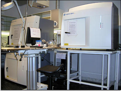
Optix MX2 Time-Domain Optical Imager
Traditional in vivo optical molecular imaging systems grossly measure all of the photons that propagate through tissue without any temporal discrimination. This is known as the Continuous Wave (CW) technique. The method is limited to providing the attenuation or the total loss of photons in tissue. It cannot discriminate absorption events from scattering events, which impedes its capability to uncouple location (depth) from concentration in the image.
 and (G)...")
Fluorescence decoupling of GFP and autofluorescence for homogenous phantom of 2 x 10⁶ NB4-GFP cells immersed in 1% liposin containing 50 ppm Indian ink at depths 0.6 mm - 6.2 mm (A-F) and (G) heterogeneous phantom of 5 x 10⁶ NB4-GFP cells placed at various anatomical locations. (A) Fluorescence images are obtained for GFP cells at 4.7 mm in liposin solution (Raw Intensity) and liposin solution only (background) and plots of their respective fluorescence lifetimes shown in (B). By subtraction of background lifetime from raw lifetime, on a pixel per pixel basis, the subtracted lifetime curve (C) of predominantly GFP vs. background fluorescence lifetime is created. A clear difference in rate of fluorescence decay and in tFDmax (inset C, used to estimate depth GFP inclusion) is observed between the GFP and background peaks. By fitting the decay tail of the of measured signals (C) using the Levenberg-Marquet least square method, the fluorescence lifetimes are obtained. Once the fluorescence lifetime is known (GFP = 2.7 ns), it can be used to gate for GFP fluorescence lifetime only and, subsequently, intensity of GFP only (D). GFP fluorescence intensity of 2x10⁶ NB4-GFP cells was decoupled from liposin autofluorescence at depths 0.6-6.2 mm analogously. Similarly, 5x10⁶ NB4-GFP cells placed at various anatomical locations in a dead mouse (F) could be decoupled from autofluorescence using the mouse autofluorescence as background. Note: Raw intensity photon counts (PC) in are not normalised. NC; normalised counts. S.C; subcutaneous, I.P; intraperitoneal, I.M; intramuscular, between the posteriolateral abdominal wall and kidney (K), under the skull (B) and within the thorax between the lungs and ribs (T).
Hovedinnhold
The imager is located at the Lab Animal Facility, but another core facility, MIC, is responsible for operations. Read more here.
18.02.2014