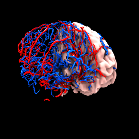
The measurement of perfusion and filtration are important clinical parameters used in diagnosis, follow-up, and therapy.
The aim of this project is to investigate a novel approach for the interpretation of dynamic medical imaging with emphasis on blood distribution and flow. Typical applications include characterisation of strokes and planning and evaluation of cancer treatment.
Medical image acquisition techniques like computerised tomography (CT), magnetic resonance imaging (MRI), or positron emission tomography (PET), can all be applied in a dynamic setting where the evolving distribution ofan injected contrast agent is gathered together as a temporal sequence of images. Quantitative tissue characterisation (e.g. blood perfusion) from such data is currently performed locally by applying tracer-kinetic methodology to a single region of interest (ROI) or a voxel at a time.
By modelling the flow from first principles and calibrate the models to observations via systematic assimilation techniques, our goal is to advance understanding and clinical utility of dynamic imaging interpretation.
In addition, this research aims to produce knowledge and technology that contributes to ICT solutions for enhancing productivity and efficiency within the health sector.