Immunohistochemistry
Immunohistochemistry requires a piece of tissue usually taken with a biopsy. The tissue sample is sliced extremely thinly and the slices (sections) are fixed on a glass slide.
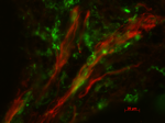
 and macrophages (green) in the root end of a molar tooth.")
Immunofluorescense image of lymphatic vessels (red) and macrophages (green) in the root end of a molar tooth.
Main content
The tissue sample has characteristic markers, or antigens, which can be used to identify the existence and localization of cells, structures etc.. Antibodies against these characteristic antigens are added on the glass slide with the tissue sections, and the antibodies bind wherever the antigens are present. Excess antibody is then washed away. The bound antibodies have labels on them that either fluoresce (glow) or undergo a chemical reaction that makes them visible under the microscope.
05.03.2020