Image processing of brains
Can mathematics help to understand how the brain functions and how it changes with age?
Main content
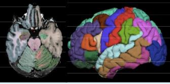
In connection with an ageing project, we have large quantity of brain MRI data sets, which have been taken of a group of people from 5 years ago and from last year. The aim is to study how the brain changes with age and compare the data sets from then and now. To do this in a systematic manner requires both registration (to compensate for, e.g., movement or misalignment) and segmentation (to focus on the area of interest).
In addition, in the past year we have received (from the same patients) data in other image modalities: diffusion-tensor images, fMRI images and perfusion images.
fMRI - functional Magnetic Resonance Imaging - is a modern imaging technique that can be used to depict the blood flow to the brain, for example, while the subject solves different tasks, and these measurements can be used to reconstruct the active areas of the brain and thereby understand how the brain works.
It is also possible to combine this data with DTI images (Diffusion Tensor Imaging), which can be used to reconstruct the nerve bundles (highways for neurological signals) in brains.
A question of interest is to see if these areas, which are active under certain stimuli (the areas may be located in different regions of the brain), are connected to each other through nerve fibres.
Perfusion images study blood-perfusion with the help of a contrast agent that appears stronger in the MR images. Then one can generate time-curves (pointwise) which can be studied (e.g. grouping by shape, intensity, slope, etc.) and they can be used for parameter estimation. E.g. for a brain with a tumour, one will typically have curves that have different profiles than other curves.
Contact: Arvid Lundervold, Antonella Z. Munthe-Kaas.
