Research from home
On Thursday 12th of March, the University of Bergen was shut down on short notice. Employees and students left their study and workplaces, worried about the recently declared pandemic situation and how the next weeks and months would be like.

Main content
The last hours before lockdown - securing precious biological material
Ongoing research projects in the laboratories were abruptly terminated with a few hours' notice. In the time before the closure, there had been a lot of activity in the lab and the master's students were in the middle of the important last lab phase before the spring master's exam.
Hanne Øye, a master's student in the NAT group, Department of Biomedicine, had spent a lot of time prior to the lockdown to modify and cultivate several valuable cell lines from cancer patients. She had recently made the latest cell cultures thrive and was ready to use these in experiments that would contribute to important results in her master's project. "With only two hours' notice, we were supposed to end all lab activities and this resulted in frantic activity to save my 11 precious cell lines," Hanne says. "The next few days at home I was worrying whether I would be able to deliver enough results for the Master's dissertation."
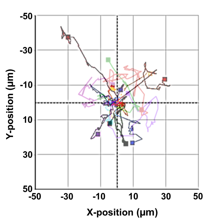
Methodological advance for studying cancer cell migration
Prior to the lockdown, Hanne, Henriette Aksnes (supervisor and researcher) and I had submitted a manuscript for review to the journal Scientific Reports. We wanted to publish a method we had used to study the invasive properties of cancer cells. This method is based on a microscopy technique for living cell cultures, called digital holographic microscopy (DHM) which we had previously used to study the migratory capacity and cell morphology of cancer cells. We now wanted to monitor the invasive potential of cancer cells and therefore investigated whether the HoloMonitor microscope managed to take good pictures for quantitative analysis when we let the cells invade through a thick layer of Matrigel (3D matrix). When we did this, we were happy to find that it worked given that it was not a listed application for the instrument. The reason for this is that the HoloMonitor is mainly developed to study cell migration in a simplified environment in the cell dish. We therefore wanted to share our experience with other cell biologists, showing that the HoloMonitor also proves to be well suited to study the invasion of cancer cells. Thus, we documented the method and put together a manuscript for an article.
Studying cells without cell lab?
A few weeks after the closure, we received the peer review report for our manuscript. The reviewers were unfortunately not completely convinced. However, they provided us with good input on how we could improve our manuscript and recommended that some of our claims should be strengthened with further data. The labs were still closed, and no additional experiments could be performed. Thus, the solutions had to come from our respective home offices. And here, the HoloMonitor's built-in software solution showed its strength with a variety of applications and gave me the opportunity to re-analyze previous image material from home. New discoveries were made that strengthened our previous results. To fully address the questions that had been raised in the peer review, I went back to my master's thesis that I submitted last year. There, I extracted data from holographic images and used fluorescence images taken with the Dragonfly system at the instrument park at the Molecular Imaging Center (MIC).
Exiting home office with both a master's degree and an article
Using digital tools and various software solutions, Hanne, Henriette and I, from each of our home offices, strengthened our scientific findings and refined manuscripts and figures. Hanne's master's thesis was considered outstanding and the improved manuscript was accepted and is now published: "Exploiting the potential of commercial digital holographic microscopy by combining it with 3D matrix cell culture assays".
"Our findings extend the use of this exciting microscopy technique. This is important because by using this methodological twist, researchers can gain increased knowledge about the metastatic properties of cancer cells and how it is affected by various drugs and the cancer cells' gene expression," Henriette summarizes.
While laboratories were shut down and many projects stopped, we were lucky with the timing of the review process and the flexibility of the analysis tool. This enabled us to revise the manuscript and supplement it with additional analyzes leading to our article being accepted for publication.
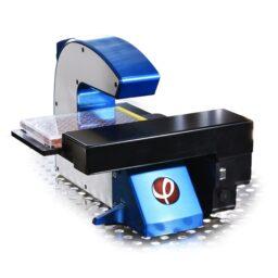
Digital holographic microscopy (DHM)
DHM generates a quantitative hologram of living cells based on how the cells material affect the microscope's laser light.
The interference pattern caused by the cells in the dish is recorded digitally. Further data processing converts the hologram into a holographic image that represents the optical imprint of morphology and 3D contour of the cells.
Source: Phase Holographic Imaging AB
Invasion