"Illuminating Life" in Nature
In the University Museum's latest exhibition, stem cell research at the University of Bergen is presented through spectacular microscopy photos. Even before the exhibition opens to the public 5 March, one of the photos can be seen on the front page of Nature's website, as the best scientific photo of the month.

Main content
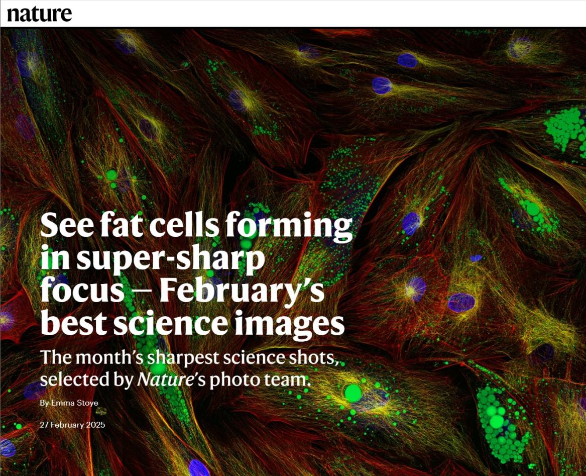
The potential in stem cells for regenerative therapies forms the basis for research conducted by postdoctoral fellow Shuntaro Yamada at the Department of Clinical Odontology at the University of Bergen. During the research process, Shuntato captures ultra-high-resolution images of cells through the microscope, illuminated and visualised with the aid of fluorescent proteins - molceules that "glow" when exposed to light on specific wavelengths. One of these images has been selected by prestigous academic journal Nature as best scientific photo of the month. As of this writing, it is also the front page image of Nature's website.
Good things come in threes
It is not the first time Shuntaro has been featured in Nature. Twice before, the acclaimed scientific journal, counted as one of the world's premier, has selected Shuntaro's scientific images as best of the month. This time, Shuntaro himself says it is his best and most detailed image he has captured so far.
In the photo, bone marrow stem cells are shown during the process of differentiating into specialised adipose tissue cells. Understanding stem cell differentiating could help researchers unclock the unique properties in stem cells, which would have huge implications for future therapies, especially where different kinds of tissue has been damaged or destroyed.
Experience this photo, and a selection of Shuntaro's other images visualising the almost otherwordly depictions of cellular processes in the the exhibition "Illuminating Life", which opens to the public 5 March 2025, at the University Museum.
Illuminated Cells:
The exhibition "Illuminating Life" presents stem cell research at UiB through Shuntaro Yamada's ultra-high-resolution microscopy images.
The exhibition is a collaboration between:
The University Museum
The Department of Clinical Odontology
Division of Research and Innovation
Bergen Tissue Engineering
Mohn Centre for Regenerative Medicine