Hyperion - Image mass cytometry
Main content
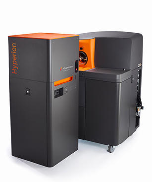
The HyperionTM Imaging System is a high-parameter imaging system capable of analyzing 4 to 37 protein markers at subcellular resolution in fixed tissue sections or cell smears. With the ability to utilize up to 135 channels to detect additional parameters, the Hyperion Imaging System is ideal to meet researcher needs today and well into the future.
The Hyperion Imaging System is based on the HeliosTM mass cytometry platform. This high-dimensional imaging platform uses mass-tagged antibodies to markers alongside cell structural features in tissue and cell smears.
Mass-tagging involves separation of signals based on the differences in mass, resulting in distinct signals for each marker without the need for compensation associated with fluorometric techniques yet far more specific and sensitive than tag-free techniques.
The metal tags can be defined within 1 Da spatial resolution in tissue and cell smears, resulting in a unique spatial and parametric definition of the cells in situ. The system enables understanding of protein behavior and interactions to drive biological breakthroughs.
Image from Hyperion project in prof. Daniela Elena Costea`s group. The project is led by Ph.d. candidate Stian Tornaas and the image is displaying the following markers: Fibroblast activation protein (FAP) = green, Podoplanin = blue, Caveolin = red, and E-cadherin = cyan

Hyperion Imaging System
| Channels | 135 |
|---|---|
| Mass range | 75–209 amu |
| Abundance sensitivity | 0.3% for 159Tb |
| Frequency | 200 pixels/sec |
| Detection limit | ≥400 copies per μm2 |
| Dynamic range | 4 orders of magnitude |
| Calibration | Automated |
| Operating system | Windows® 7 Pro 64-bit |
| Data storage | >6 TB RAID (mirrored) |
| Switch time (between modes) | 12 hr |
| Cross-cell contamination (redeposition) | ≤2% (selected direction) |
| Crosstalk pixel to pixel | ≤15% at 200 pixels/sec |
| Wet tissue thickness for full ablation | ≤7 μm |
| Addressable sample size on slide | ≥15 mm x 45 mm |
| Optical view of sample | ≥ 250 μm x 250 μm |
| File type | TXT, multipage TIFF, OME-TIFF, MCD |
| Scan area | ≥1 mm2/2 hr (@200 Hz) |
https://www.fluidigm.com/products/hyperion-imaging-system#specifications
The Hyperion image mass cytometer was funded by the Faculty of Medicine, the Haukeland University Hospital and the Center for Cancer biomarkers (CCBIO).
An antibody reagent repository has been purchased and aliquotes of antibodies can be purchased though the booking system. Meaning that core facility will provide/sell panels to researches and groups that would like to test and use image mass cytometry. This will allow researches and groups to more easily test mass cytometry on their own samples. This will give then better understanding of what they can do and they can get their hands on some data on their specific samples. See list of antibodies below
Human Immuno-Oncology IMC Panel, 31 Antibodies (201509) Cost: 200,-/ul
Panel can also be found at the bottom of the page as a pdf


Conjugation kits can also be purchased though the core, see https://www.uib.no/en/clin2/flow/102286/cytofxt-helios


The Hyperion is located on 5th floor of the Laboratory Building at UiB. Access and management of this platform is through the Flow Cytometry Core Facility